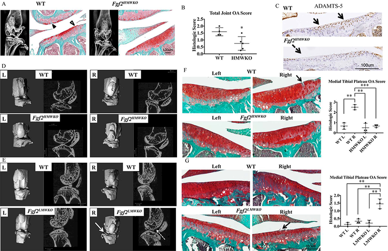
Figure 5. Fgf2HMWKO are protected from spontaneous (females) and mechanically-induced (males) OA.

A, 2 year old Fgf2HMWKO and WT have similar subchondral bone phenotypes as determined by x-ray. Safranin-O staining of lateral side of joint shows loss of proteoglycan content (closed arrowhead) and fibrillation of cartilage (open arrowhead), compared to the more robust proteoglycan content of Fgf2HMWKO samples. N=4–6/group. B, Mean total joint OA score of Fgf2HMWKO is significantly lower than WT. Values are means ± SD, n=4–5/group. C, Representative immunohistochemical staining shows decreased protein expression of ADAMTS-5 along tibial articular cartilage of Fgf2HMWKO (dashed arrow) compared to expression in WT littermates (arrows). D-E, 3D reconstructed images and μCT images of sagittal sections of right tibial-loaded knees (2 weeks after loading) of 21 month old Fgf2HMWKO mice appear to have a similar appearance to the left contralateral control and both left and right knees of WT. Right tibial loaded knees of Fgf2LMWKO mice show erosion of the tibial surface (arrow) compared to the left contralateral control and all samples in D. Sagittal images display decreased femoral subchondral bone (dashed arrow) and increased trabecular spacing and decreased trabecular number in epiphyses compared to the contralateral control n=3–4/group. F, Safranin-O staining shows fissures (arrow) in cartilage of right tibial-loaded knees (2 weeks after loading) of 21 months old WT littermates of Fgf2HMWKO mice. OA score of right medial tibia of WT littermates of Fgf2HMWKO mice was significantly higher than all others. G, Right tibial loaded knees of 12 week old Fgf2LMWKO mice (2 weeks after loading) show fraying (arrow) and a significantly higher OA score than controls. Values are means ± SD, p <0.05**p <0.01, ***p <0.001.
