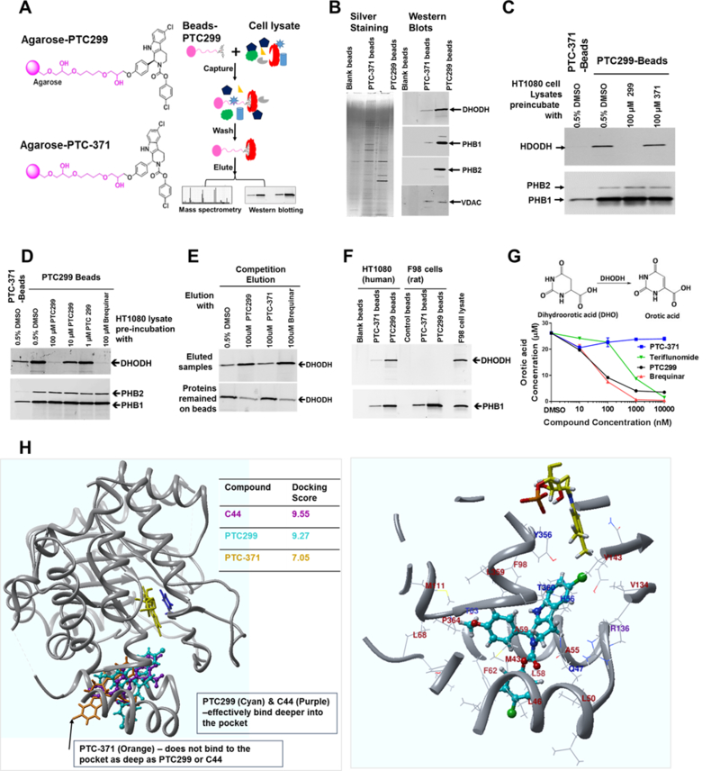
Figure 3. PTC299 binds to and inhibits the activity of DHODH.

(A) Schematic drawing of PTC299, the inactive isomer linked to the agarose beads and the outline of chemical pulldown study; (B) PTC299 beads bind to DHODH and prohibitin-1/2. Pull-down samples from Blank, PTC-371 and PTC299 conjugated beads were resolved on 4–20% SDS-PAGE. Left panel shows the silver staining, while the right panel shows westerns blotted with antibodies against human DHODH, PHB1 (prohibitin-1), PHB2 (prohibitin-2) and VDAC (voltage-dependent anion channel) respectively; (C) PTC299 selectively inhibits DHODH binding to the PTC299-beads. HT1080 cell lysates were preincubated with 100 μM free PTC299 or PTC-371 as indicated in the figure for 2 hours at 4oC prior to the pull-down study described in the methods; (D) Western blot analysis demonstrated that free PTC299 and brequinar competes DHODH binding to the PTC299-beads. The experiment was done in the same way as for Fig.3C. (E) Western blot analysis demonstrated that free PTC299 selectively elutes DHODH from the PTC299-beads; (F) PTC299 conjugated beads selective bind to human rather than rat DHODH; (G) PTC299 inhibits DHODH activity in isolated mitochondria, data are representative of three independent mitochondria preparations, orotate production was quantified by LC-MS/MS analysis of the end product after 30 minutes enzyme reaction. (H) Computer modelling using crystal structures of the human DHODH protein. Both the brequinar analog C44 (Purple) and PTC299 (cyan) bind deep in the pocket, but the inactive enantiomer PTC-371 (orange) does not, which is reflected by the docking scores on the right. The cofactor flavin mononucleotide (FMN) and the product orotate are shown in yellow and blue respectively. Right panel shows a localized view that PTC299 (cyan) binds to the pocket of human DHODH crystal at 4IGH. Amino acids within 5 angstroms of PTC299 are shown as a ribbon. Amino acid residues within 3 angstroms were labeled (hydrophobic, brown; charged, purple; polar, blue). Relevant side chains that make up the pocket are displayed explicitly.
