Fig. 4:

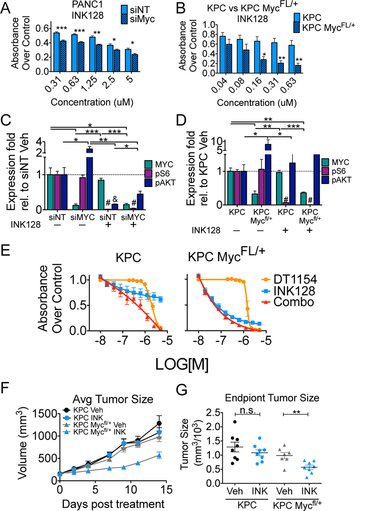
Decreased Myc expression sensitizes PDA cells to mTOR inhibition in vitro and in vivo A. PANC1 cells transfected with non-targeting siRNA (siNT) or MYC targeting siRNA (siMYC) were treated with increasing concentrations of INK128. Viability was assessed by MTS 72 hours after drug treatment. Mean +/−SEM of three biological replicates. B. Cells derived from KPC or KPC Mfl/+ mice were treated with increasing concentrations of INK128. Viability was assessed by MTS 72 hours after drug treatment. Mean +/−SEM of three biological replicates. C. Cells from panel A (siNT or siMYC) and D. cells from panel B (KPC or KPC Mfl/+) were treated with either Vehicle or 0.5μM INK128 for 6 hours. Lysates were analyzed by western blot for MYC, pAKT (S473), pS6 (S235/6) and totals. Quantification from three biological replicates is shown. Mean +/−SEM. E. KPC (left) and KPC Mfl/+ (right) cells were treated with either Vehicle or increasing concentrations of DT1154, INK128, or the combination for 72 hours. Viability relative to Vehicle was assessed by MTS. Average of 4 replicates across 2 biological experiments shown. Mean +/−SEM. For all panels ***p<0.001 **p<0.01 *p<0.05 by two-tailed students t-test. #, & denotes pS6 or pAKT levels, respectively, are significantly different in INK128 conditions compared to Vehicle. F. KPC and KPC Mfl/+ cells were injected subcutaneous and then treated with either Vehicle or INK128 (0.5 mg/kg oral gavage, once a day/6 days a week). Tumor volume was measured across time. Mean +/− SEM. n= 8 KPC, 8 KPC+INK128, 7 KPC Mfl/+, 8 KPC Mfl/++INK128. G. Quantification of end point tumor size from panel F. **p<0.01 by two-tailed students t-test
