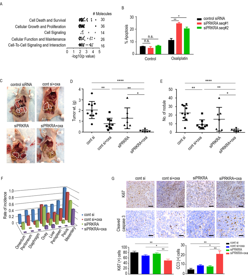
Figure 2.

Anti-tumor effects of the combination of oxaliplatin and siPRKRA. A, The five molecular functions most significantly altered as a result of downregulated mRNAs under PRKRA-silenced conditions. Data are from RNA-sequencing of cells transfected with siPRKRA or control siRNA. The data were subjected to Ingenuity Pathway Network analysis. B, Apoptosis assay for oxaliplatin and siPRKRA in RMUG-L-ip1 cells at the end of 72-hour time point. Cells were transfected 6 hours and 48 hours after plating. Chemotherapy treatment was started 72 hours after plating the cells. The oxaliplatin dose used for the assay was 1 μM. C, Representative images of the extent of metastatic spread of MOC in mice given a control siRNA, control siRNA plus oxaliplatin, siPRKRA, or siPRKRA plus oxaliplatin (n = 10 per group). D and E, tumor weights (D) and nodule numbers (E) in each mouse group 43 days after RMUG-L-ip1 cell injection. F, Distribution of metastatic nodules in each mouse group. G, Immunohistochemical staining of MOC samples obtained from RMUG-L-ip1 orthotopic models for Ki67 and caspase 3. Scale bar=100μm. The in vitro data are presented as means ± standard error of the mean for at least three experimental groups. *P < 0.05; **P < 0.01; ****P < 0.0001 (Student t-test).
