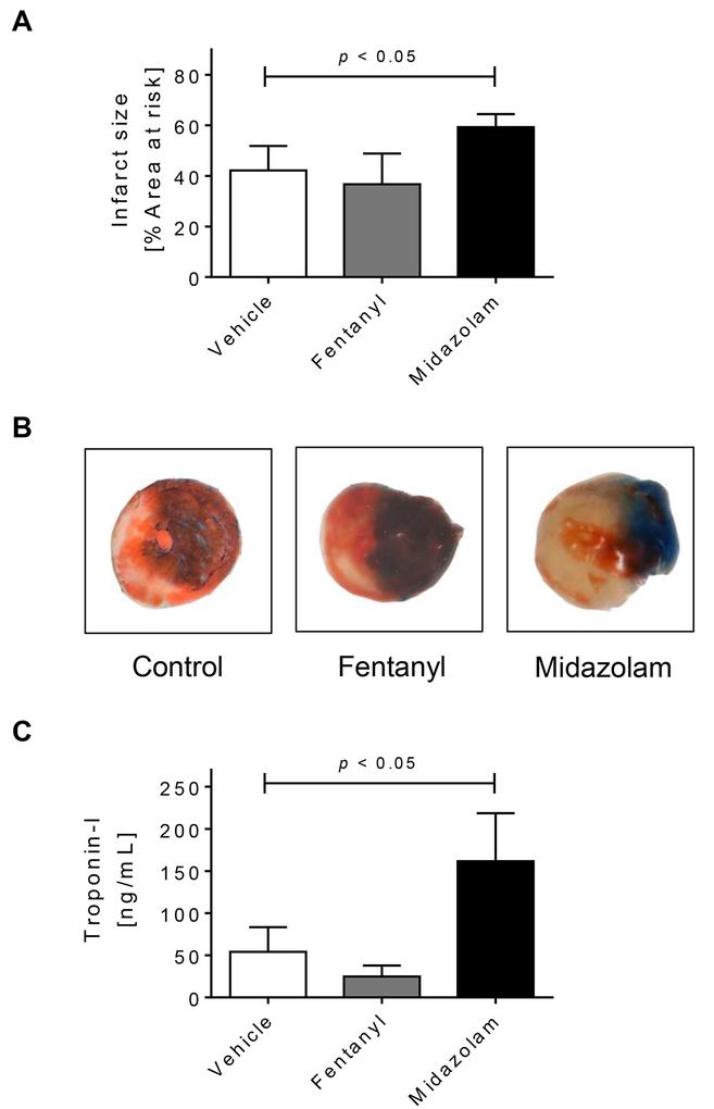
Figure 2. Midazolam in myocardial ischemia and reperfusion injury.
Mice were pretreated with vehicle (NaCl 0.9%), fentanyl (1mg/kg) or midazolam (200mg/kg) i.p. 2 hours prior to myocardial ischemia. Myocardial ischemia consisted of 60 min of ischemia followed by 120 minutes of reperfusion. Infarct sizes were measured by double staining with Evan’s blue and triphenyl-tetrazolium chloride. Infarct sizes are expressed as the percent of the area at risk (AAR) that underwent infarction. Serum troponin I concentrations were measured by enzyme-linked immunosorbent assay (ELISA). (A) Infarct sizes as the percent of AAR; (B) Representative infarct staining; (C) Serum troponin I concentrations; (D) Linear Regression analysis between Per2 transcript and troponin I levels; (n=5–7; mean±SD; p<0.05).

