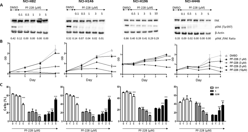
Figure 1: PF-573,228 (PF-228)’s effect on FAK expression/activity, cell proliferation, and cell cycle in SCLC cell lines.

A. FAK expression and phosphorylation evaluation by Western blot (WB). Whole cell lysates from four SCLC cell lines treated with PF-228 or DMSO control for 90 min. were resolved by sodium dodecylsulfate-polyacrylamide gel electrophoresis (SDS-PAGE) and blots were incubated with antibodies against total FAK (125 kD), phospho-FAK (Tyr397) (125 kD), and β-Actin (45 kD) for normalization. Dose-dependent inhibition of FAK phosphorylation (Tyr397) was observed by WB in cell lines treated with PF-228 as compared to those treated with DMSO, while total FAK expression was not modified. WB densitometric quantification is available in Supplementary Fig.S1. B. Cell proliferation evaluation by WST-1 assay. Four SCLC cell lines were cultured for three or four days in presence of PF-228 or DMSO. Dose-dependent inhibition of proliferation was observed by WST-1 assay in cells treated with PF-228 as compared to those treated with DMSO. Optical density (OD) in Y-axis reflects the proportion of metabolically active cells. Error bars represent mean +/− standard deviation (SD) (n=3). All the graphs represent one of three independent experiments with similar results. *** P ≤ 0.001. C. Cell cycle evaluation by flow cytometry. Four SCLC cell lines treated with PF-228 or DMSO for 24h were stained with anti-BrdU antibody and propidium iodide (PI), and the staining was quantified by fluorescence-activated cell sorting (FACS) analysis. Dose-dependent inhibition of DNA synthesis and induction of cell cycle arrest in G2/M phase was observed by flow cytometry in cell lines treated with PF-228 as compared to those treated with DMSO. Error bars represent mean +/− SD from three independent experiments. *** P ≤ 0.001.
