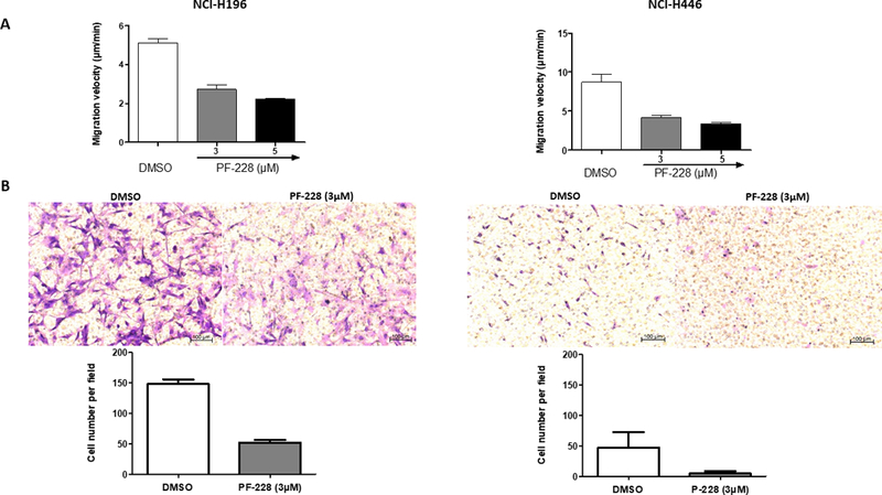
Figure 3: PF-228’s effect on migration and invasion in SCLC cell lines.

A. Migration evaluation by wound healing assay associated with time-lapse microscopy. Two adherent SCLC cell lines were grown to confluence, wounded, incubated overnight with culture medium, and then treated with PF-228 or DMSO for 12h. Cells were monitored during these 12h using a Zeiss Axiovert 200M microscope (Zeiss, Thornwood, NY). Images were captured every 15min. Velocity of cell migration was measured using ImageJ. Decreased motility was observed in cell lines treated with PF-228 as compared to those treated with DMSO. Error bars represent mean +/− SD from three independent experiments. B. Invasion evaluation by modified Boyden Chamber assay. Two adherent SCLC cell lines (one adherent and one with mixed-morphology) were seeded on the top of an insert pre-coated with matrigel and separating the two chambers of a transwell. Culture medium containing 1%-FBS was placed in the upper chamber and 10%-FBS in the lower chamber. After 12h-treatment with PF-228 or DMSO, cells that moved through the pores towards the bottom of the insert were stained with crystal violet, digitally pictured, and quantified by the free software ImageJ (NIH, Bethesda, MD, USA). Right panels are pictures of SCLC cell lines stained with crystal violet on the lower side of the insert which are representative of the numerous fields (x10 magnification) analyzed in two independent experiments performed in duplicate wells. Left panels represent quantification of the number of cells per field on the bottom of the insert. Decreased invasion was observed in cell lines treated with PF-228 for 12h as compared to those treated with DMSO. Error bars represent mean +/− SD from two independent experiments.
