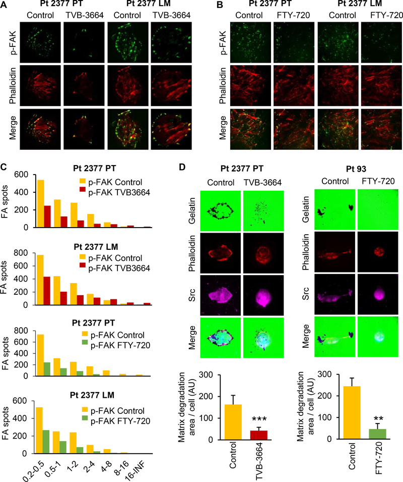
Figure 4. Inhibition of FASN and sphingolipid metabolism diminishes formation of focal adhesion complexes.
Primary Pt 2377 PT and Pt 2377 LM CRC cells were treated with (A) 0.2 μM TVB-3664 for 7 days and (B) 2.5 μM FTY-720 for 48h, stained with focal adhesion marker p-FAK (Y397) antibody (green) and phalloidin (red) and images were captured using TIRF microscopy. Representative images are shown. (C) Number of focal adhesions in different size classes was quantified using NIS-Elements. (D) Primary Pt 2377 PT cells were treated with 0.2 μM TVB-3664 for 7 days. Primary Pt 93 CRC cells were treated with 2.5 μM FTY-720 for 48h. Gelatin degradation ability of cells was assessed. Background gelatin is stained with Alexa Fluor 488 (green). Graphs display quantification of gelatin degradation per cell in both control and treated cells.

