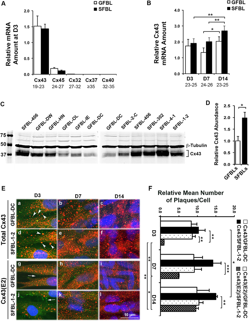
Fig. 2. Cx43 expression and localization in cultured gingival and skin fibroblasts.
(A) Results show qPCR analysis of relative mRNA amount of major Cxs in gingival (GFBLs; GFBL-DW, GFBL-HN, GFBL-OL, GFBL-IE, and GFBL-DC) and skin (SFBLs; SFBL-2-C, SFBL-406, SFBL-302, SFBL-4–1, and SFBL-1–2) fibroblast cultures at day-3 post-seeding. Cx43 was the major Cx expressed in both GFBLs and SFBLs. (B) qPCR analysis of relative Cx43 mRNA amount in GFBLs and SFBLs, 3-, 7-, and 14-days post-seeding. Results represent mean ± s.e.m. from a minimum of three repeated experiments (* p < 0.05, ** p < 0.01; two-tailed Student’s t-test). Results in A and B represent mRNA amount relative to GFBL-DC. Range of the Ct-values obtained from qPCR is indicated below each gene name. (C and D) Western blotting analysis (C) and quantitation (D) of Cx43 in GFBLs and SFBLs day-7 (D7) 3D cultures. SFBLs showed significantly higher abundance of Cx43 compared to GFBLs. Sample loading was normalized for β-Tubulin levels. (E) Representative images from GFBL-DC and SFBL-1–2 day-7 3D cultures double-immunostained for total Cx43 (red) and ZO-1 (green; indicator of cell-cell contacts) (a–f), or HC-associated Cx43 (Cx43(E2); red) and ZO-1 (g–l). (a–f) Cx43 staining was abundantly present in both GFBLs and SFBLs throughout the cell body. In addition, some Cx43 staining colocalized with ZO-1 staining at cell-cell contact areas (arrowheads in a and d), likely representing GJ plaques. (g–l) GFBLs and SFBLs also showed numerous Cx43(E2)-positive plaques that did not colocalize with ZO-1 (arrows in g and j) at the cell-cell contacts, likely representing Cx43 HCs. Nuclear staining (blue) was performed using DAPI. Representative immunostaining images from three parallel samples from two repeated experiments are shown. (F) Quantitation of mean number of total and HC-associated Cx43 plaques per cell over time in culture in GFBL-DC and SFBL-1–2. For quantitation, five standard fields from each coverslip were randomly selected. Cx43-positive plaques were counted in minimum of 100 cells per field. Results are relative to the number of Cx43(E2)-positive plaques in SFBL-1–2 at day-3 post-seeding.

