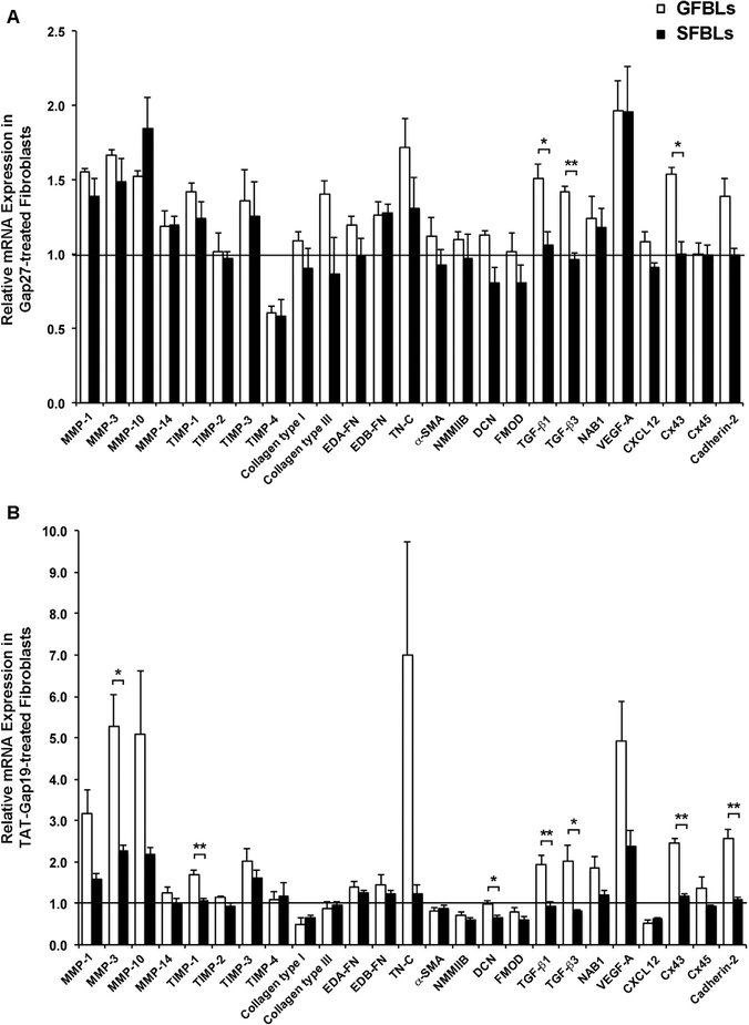
Fig. 5. Gene expression response to Gap27 or TAT-Gap19 treatment in human gingival and skin fibroblast cultures.
Day-7 3D cultures of GFBLs (GFBL-DC, GFBL-IE, and GFBL-DW) and SFBLs (SFBL-1–2, SFBL-4–1, and SFBL-302) were treated with (A) Gap27 or control peptide (150 μM), or (B) TAT-Gap19 or control peptide (400 μM) for 24 h, and the expression of a set of genes involved in wound healing was analyzed by qPCR. Results represent mean ± s.e.m. of amount of mRNA relative to control peptide-treated cells ( = 1). Statistical testing was performed between GFBLs and SFBLs for each peptide treatment (* p < 0.05, ** p < 0.01; two-tailed Student’s t-test). Horizontal line ( = 1) indicates relative amount of mRNA for the control peptide-treated samples in both GFBLs and SFBLs. EDA-FN: Extra Domain A-Fibronectin; EDB-FN: Extra Domain B-Fibronectin; TN-C: Tenascin-C; α-SMA: α-Smooth Muscle Actin; NMMIIB: Non-Muscle Myosin IIB; DCN: Decorin; FMOD: Fibromodulin; NAB1: NGFI-A Binding Protein-1; VEGF-A: Vascular Endothelial Growth Factor-A.

