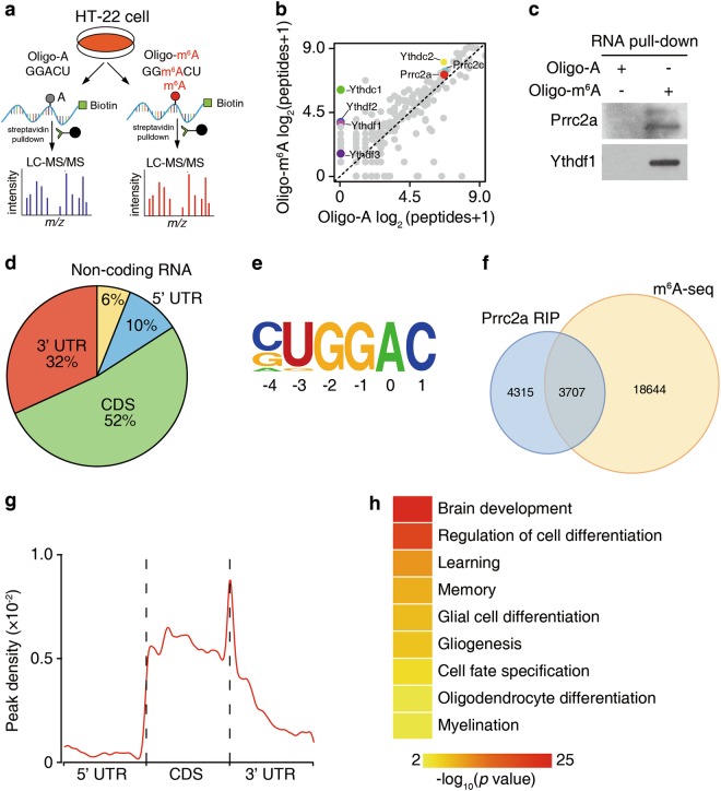
Fig. 1.
Prrc2a is a novel m6A reader. a Schematic illustration of m6A binding protein screening. b Scatter plot of proteins bound to Oligo-m6A vs Oligo-A RNA oligos. The plot was based on the average peptide numbers of proteins detected in two replicates. Enriched Prrc2a, Prrc2c, and YTH-domain containing proteins were highlighted (see also Supplementary information, Table S1). c Western blotting showing Ythdf1 and Prrc2a pulled down with an m6A-containing RNA probe. d Pie chart depicting the distribution of Prrc2a-binding peaks. e Binding motif identified by HOMER with Prrc2a-binding peaks (p = 1e-46). f Overlap of Prrc2a-binding peaks and m6A-containing peaks. g Distribution of m6A-containing Prrc2a peaks across the length of mRNA. 5′ UTR, CDS, and 3′ UTR were each binned into regions spanning 1% of their total length, and the percentages of m6A-containing Prrc2a peaks that fall within each bin were determined. The moving averages of m6A-containing Prrc2a peak percentage are shown. h Representative Gene Ontology (GO) terms of the biological process categories enriched in transcripts with both Prrc2a-binding and m6A peaks. Gene ontology (GO) analysis was performed using the DAVID bioinformatics database. GO classification for cellular component, biological process, and molecular function were performed with default settings

