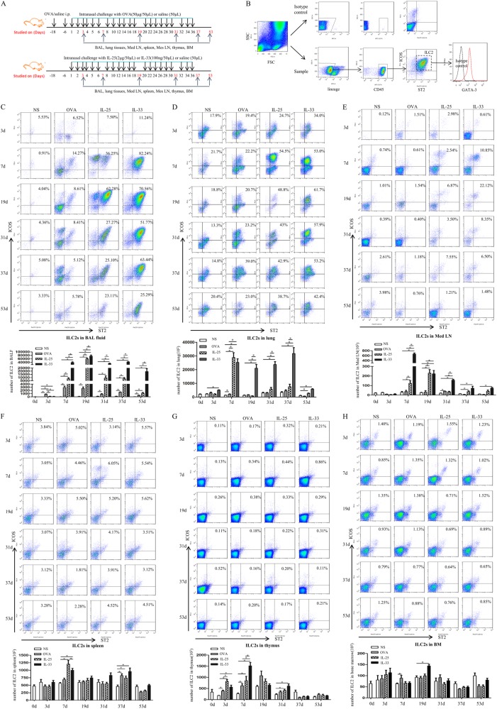
Fig. 1.
a Schedule of murine challenge. b ILC2s were gated by the protocol. (C-H) Flow cytometric identification of ILC2s in single-cell suspensions of the BAL fluid (BALF, c), lung parenchyma (d), mediastinal lymph nodes (Med LN, e), spleen (f), thymus (g) and bone marrow, (h) of treated mice. Top panels: plots showing expression of the indicated markers on lineage-negative cells (samples from 5 mice/group pooled for ILC2 staining). Bottom panels: quantification of ILC2s in treated groups. Bars show the mean ± SEM (n = 5 in each group at each time-point). *p < 0.05

