Fig. 4.
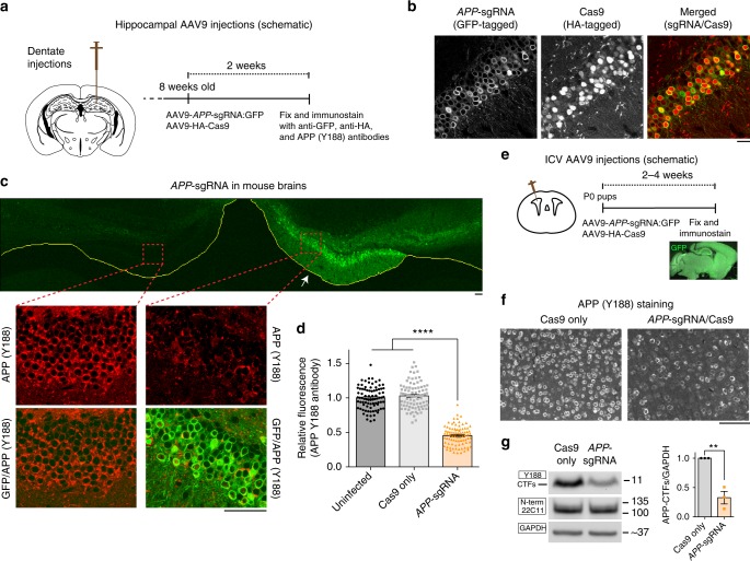
Gene editing of APP C-terminus in vivo. a AAV9-sgRNA and AAV9-Cas9 were stereotactically co-injected into dentate gyrus of 8-week old mouse brains (bottom). Two weeks after viral delivery, brains were perfused, fixed, and immunostained with anti-GFP, anti-HA and anti-APP(Y188) antibodies. b Co-expression of AAV9-sgRNA-GFP and AAV9-HA-Cas9 in the dentate gyrus. Note that majority of neurons are positive for both GFP and HA (~87% of the cells were positive for both; sampling from 3 brains). Scale bar 20 μm. c, d Coronal section of a mouse hippocampi injected on one side (marked by arrow) with the AAV viruses as described above. Note attenuated Y188 staining of neurons on the injected side, indicating APP-editing. The image of mouse hippocampus injected with Cas9 only is not shown. Fluorescence quantified in d, mean ± SEM, data from three brains. One-way ANOVA: p < 0.0001. Tukey’s multiple comparisons: p = 0.4525 (Un-injected vs. Cas9 only); p < 0.0001 (Un-injected vs. APP-sgRNA); p < 0.0001 (Cas9 only vs. APP-sgRNA). Scale bars 50 μm. e Intracerebroventrical injection of the AAV9 viruses into P0 pups. Note widespread delivery of sgRNA into brain, as evident by GFP fluorescence. Scale bar 100 μm. f Brain sections from above were immunostained with the Y188 antibody. Note attenuated Y188 staining in the APP-sgRNA/Cas9 transduced sample, suggesting APP-editing. g Western blots of the brains from e. Note decreased expression of CTFs in the APP-sgRNA/Cas9 transduced brains; blots quantified on right (mean ± SEM of three independent experiments, **p < 0.01 by two-tailed t-test). Source data are provided as a Source Data file

