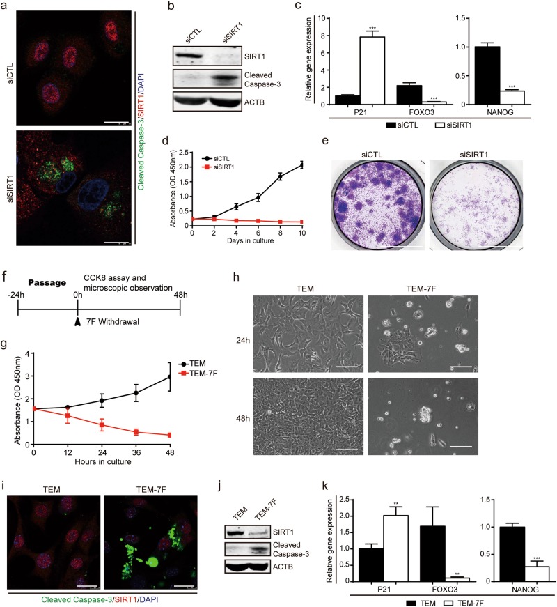
Fig. 2.
SIRT1 is essential for hepatocyte-to-LPC conversion and expansion. a Immunofluorescence images of SIRT1 and cleaved-caspase-3 at day 4 in cells transfected with control siRNA (siCTL) or siRNA-targeting SIRT1 (siSIRT1). Scale bars, 20 μm. b Western blot analysis of SIRT1 and cleaved-caspase-3 expression as in (a). c QPCR analyses for the expression of P21, FOXO3 and NANOG in cells transfected with siCTL or siSIRT1. Error bars represent s.d.; n = 3 donors (two-tailed unpaired t-test, ***P < 0.001). d CCK-8 analyses demonstrating suppression of cell proliferation in the presence of siSIRT1. Error bars represent s.d.; n = 5 technical replicates from one donor. e Light microscopy images show a reduction in clone formation following inhibition of SIRT1 by crystal violet staining. Scale bars, 10 mm. f Schematic of the 7 factors (7 F) withdrawal assay during passage. g CCK-8 analyses demonstrating suppression of cell proliferation in the absence of the 7 F within 48 h. Error bars represent s.d.; n = 5 technical replicates from one donor. h Light microscopy shows reduced cell proliferation in response to the withdrawal of the 7 F at 24 h and 48 h. Scale bars, 100 µm. i Immunofluorescence images of SIRT1 and cleaved-caspase-3 in HepLPCs cultured in TEM or TEM-7F at 24 h. Scale bars, 20μm. j Western blot analysis of SIRT1 and cleaved caspase-3 expression as in (i). k QPCR analyses for the expression of P21, FOXO3 and NANOG as in (i). Error bars represent s.d.; n = 3 donors (two-tailed unpaired t-test, **P < 0.01, ***P < 0.001)

