Fig. 6.
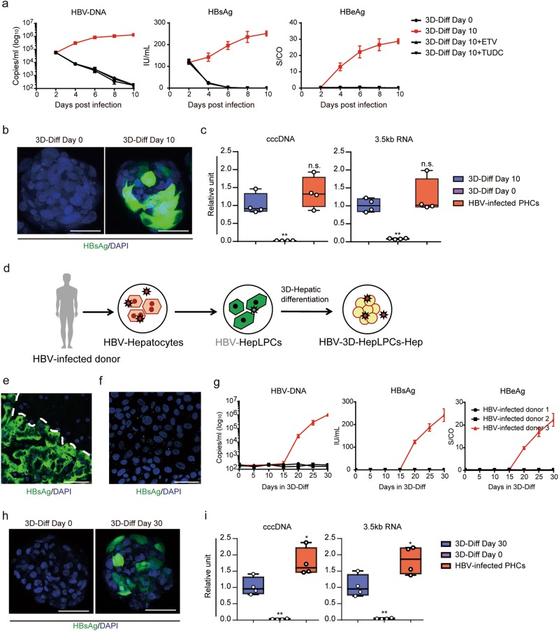
HBV infection and reactivation in 3D-HepLPCs-Hep. a Extracellular HBV-DNA and secreted viral antigens were monitored from 2 to 10 days post infection in 3D-HepLPCs treated with ETV or TUDC. Error bars represent s.d.; n = 4 donors. b HBsAg staining in 3D-HepLPCs with or without differentiation at 8 days post infection. Scale bars, 25 μm. c QPCR analyses for the expression of cccDNA and 3.5 kb RNA in 3D-HepLPCs with or without differentiation versus HBV-infected PHCs (10 days post infection, n = 4 donors, one-way ANOVA with Dunnett correction for multiple comparisons, n.s., non-significant, **P < 0.01). d Schematic of HBV reactivation in 3D-HepLPCs-Hep derived from HBV-infected donor. e HBsAg staining of the liver tissue collected from HBV-infected donor and f disappearance after 10-day culture in TEM. Scale bars, 50 μm. g Extracellular HBV-DNA and secreted viral antigens are monitored from day 0 to day 30 in HMM + DMSO in 3D-HepLPCs derived from three HBV-infected donors, n = 3 technical replicates. h HBsAg staining of patient-derived 3D-HepLPCs with or without 30-day differentiation. Scale bars, 25 μm. i QPCR analyses for the expression of cccDNA and 3.5 kb RNA in patient-derived 3D-HepLPCs with or without 30-day differentiation versus HBV-infected PHCs (10 days post infection, n = 4 technical replicates, one-way ANOVA with Dunnett correction for multiple comparisons, *P < 0.05, **P < 0.01)

