Fig. 2.
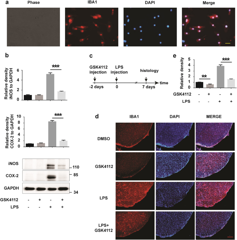
GSK4112 inhibited LPS-induced microglial activation in vivo. a Primary cultured microglia were stained using an IBA1 (red) antibody. The nuclei were stained with DAPI (1 μg/mL) (blue). Scale bars, 20 μm. b Primary cultured microglia were treated as described in Fig. 1f. Cell lysates were analyzed by Western blot for iNOS, COX-2, and GAPDH. The relative band intensities of iNOS and COX-2 to GAPDH were analyzed. ***P < 0.001, n = 3. c, d Mice received stereotaxic injection of DMSO or GSK4112 for 2 days and were then stimulated with PBS or LPS for 7 days. Mouse brains were cut into slices, and microglia were stained with an IBA1 antibody. Representative images of IBA1 (red) immunofluorescence staining in VTA are presented at AP −3.3 mm. The nuclei were stained with DAPI (1 μg/mL) (blue). Scale bar, 100 μm. e Intensity of IBA1 immunofluorescence signals in d was analyzed. **P < 0.01, ***P < 0.001, n = 3

