
Fig. 1.
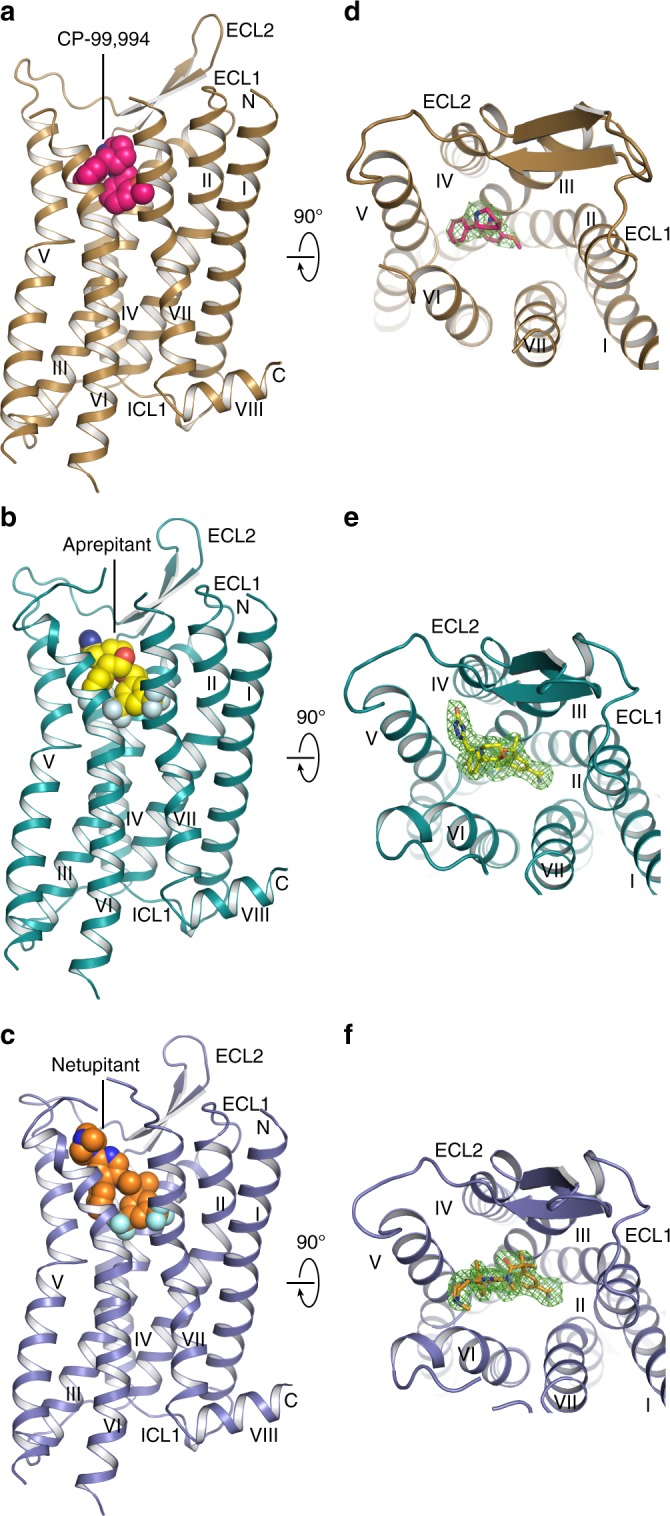
Overall structures of antagonist-bound NK1R. a–c NK1R in complex with CP-99,994 (a), aprepitant (b) and netupitant (c), viewed parallel to the membrane plane. The receptors are depicted by ribbons and coloured in brown, turquois and blue, respectively. The ligands CP-99,994, aprepitant and netupitant are shown as spheres and coloured in pink, yellow and orange, respectively. Oxygen, nitrogen and fluorine atoms of the ligands are highlighted in red, blue and grey, respectively. d–f NK1R in complex with CP-99,994 (d), aprepitant (e) and netupitant (f), viewed from the extracellular space and coloured as in (a–c). The ligands are depicted as sticks. 2Fo-Fc electron density maps of the ligands are shown in green mesh contoured at 1.0 σ