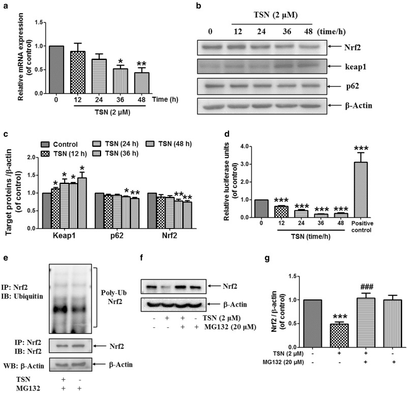
Fig. 3.
TSN reduces the expression and activation of Nrf2 in hepatocytes. a L-02 cells were incubated with TSN (2 μM) for the indicated time, and cellular mRNA expression of Nrf2 was detected (n = 3). b L-02 cells were incubated with TSN (2 μM) for the indicated time, and cellular protein expression of Nrf2, keap1, and p62 was detected. The results represent three to four independent experiments. c Quantitative densitometric analysis of Nrf2, Keap1, and p62 protein (n = 3–4). d The Nrf2/1 transcription response element-containing construct was transfected into L-02 cells. After transfection, the cells were incubated with TSN (2 μM) for the indicated time. Luciferase activities were measured using a luciferase assay system. Nrf2/1 luciferase activity was expressed as a fold induction of control cells (n = 3). e L-02 cells were pre-treated with MG132 (20 μM) for 2 h and were then incubated with TSN (2 μM) for another 36 h. Cell extracts were subjected to immunoprecipitation with anti-Nrf2 antibody, and the conjugates were detected with anti-ubiquitin and anti-Nrf2 antibodies. Each blot represents one of three independent experiments. f L-02 cells were pre-treated with MG132 (20 μM) for 2 h and were then incubated with TSN (2 μM) for another 36 h. Cellular Nrf2 expression was detected, and β-actin was used as a loading control. The results represent three repeated experiments. g Quantitative densitometric analysis of Nrf2 protein (n = 3). The data are expressed as the mean ± SEM. *P < 0.05, **P < 0.01, ***P < 0.001 vs. control; ###P < 0.001 vs. TSN

