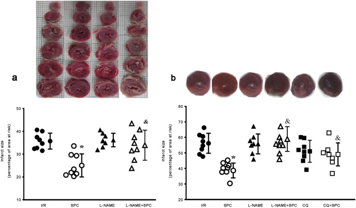
Fig. 2.
SPC-induced decreases in infarct size were blocked by l-NAME and CQ in the ex vivo and in vivo models. TTC-stained images of myocardial infarct size expressed as a percentage of the area at risk (a, ex vivo and b, in vivo). Anesthetized rats underwent 30 min of ischemia followed by 2 h of reperfusion (I/R) with or without sevoflurane treatment Means ± SD. *P < 0.05 vs. I/R group; &P < 0.05 vs. SPC group (n = 8/group)

