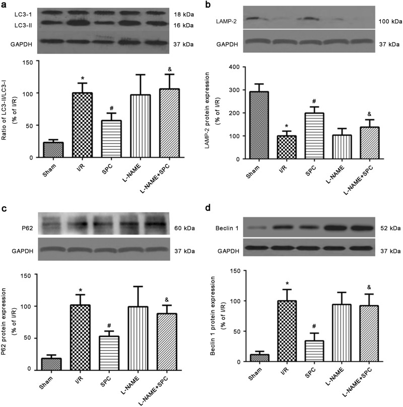
Fig. 8.
SPC-enhanced autophagic flux was blocked by the NOS inhibitor l-NAME in ex vivo experiments. a Immunoblots and densitometric analysis of LC3-I and LC3-II in heart tissues. b Immunoblots and densitometric analysis of LAMP-2 in heart tissues. c Immunoblots and densitometric analysis of P62 in heart tissues. d Immunoblots and densitometric analysis of Beclin 1 in heart tissues. The data are presented as the means ± SD. *P < 0.05 vs. Sham group; #P < 0.05 vs. I/R group; &P < 0.05 vs. SPC group (n = 5 hearts/group)

