
Fig. 5.
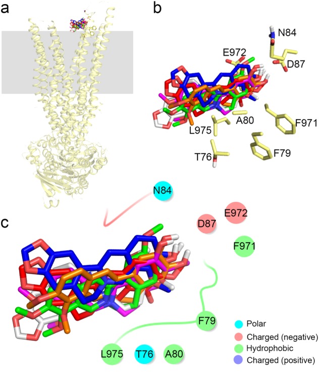
The binding docking results of the six compounds to P-gp in the release stage (Z ~ 27Å). Membranes are shown as gray squares, and the binding modes of the six compounds are shown (a). Amino acids within 4 Å of the compounds are shown as yellow sticks (b). Interactions of BBR, BER, COL, JAT, THA, and DEM with P-gp (c)