Figure 1.
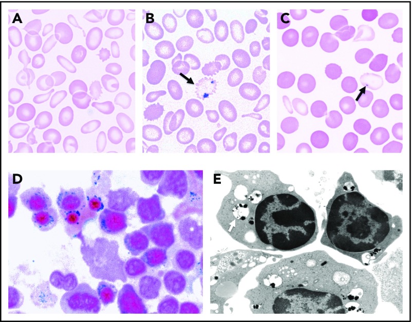
Morphological features of SA. (A) May-Gruenwald-Giemsa (MGG)-stained peripheral blood smear of a man with mild XLSA demonstrating hypochromia, anisocytosis, and microcytosis. (B) Iron-stained peripheral blood smear from the same patient highlighting a siderocyte (arrow). (C) MGG-stained peripheral blood smear from the patient’s mother demonstrating the dimorphic red blood cell population, including hypochromic microcytes containing Pappenheimer bodies (arrow). (D) Iron-stained bone marrow aspirate smear from a man with XLSA demonstrating iron granules (blue) ringing around late erythroblast nuclei. (E) Transmission electron micrograph from a patient with RARS demonstrating electron densities (black) within degenerating mitochondria (pale vacuoles, indicated by an arrow) ringing around erythroblast nuclei (photograph courtesy of Marcel Seiler, Boston Veterans Affairs Medical Center [VAMC], Boston, MA). (A-D) Equally scaled; original magnification ×1000. (E) Original magnification ∼×8000.

