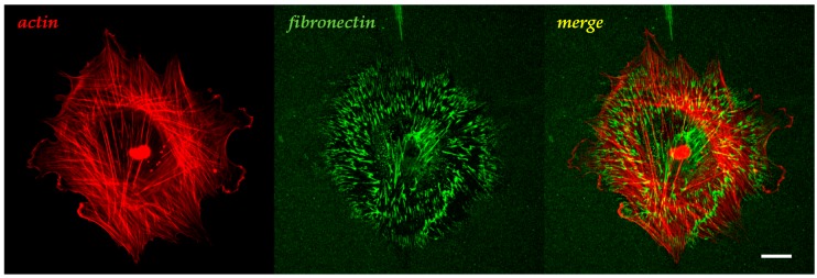
Figure 1.
Confocal micrograph showing the effect of cell-generated forces on physisorbed fibronectin. MC3T3 preosteoblasts cultivated for 12 h on a nanograted, O2 plasma-treated PDMS substrate. Fibronectin (10 µm/mL) undergoes extensive remodelling caused by contractile forces. Fibronectin compaction is observed at both ends of actin fibers. Note how fibronectin smears follow the actin direction and leave a dark halo upon compaction. Actin is stained with Tetramethylrhodamine B isothiocyanate-phalloidin (red); fibronectin is stained by immunofluorescence (green). Scale bar: 20 µm.

