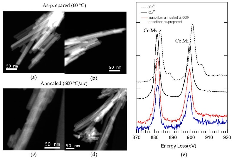
Figure 13.
(a–d) STEM-HAADF images of C12GA-templated CePO4 sample, as-prepared (a and b) and annealed at 600 °C/air (c and d); (e) Corresponding EELS spectra performed in a nanofiber of as-prepared and 600 °C-annealed templated CePO4; For comparison purposes, the electron energy loss near edge structure (ELNES) spectra of Ce3+ and Ce4+ ions are also shown as a reference [102].

