Figure 1.
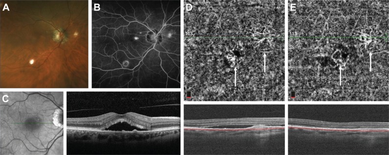
Fundus photograph of the right eye with POHS showing peripapillary atrophy and multiple “punched out” chorioretinal scars (A). Late-phase fluorescein angiogram demonstrating a small area of hyperfluorescence at the fovea (B). Structural OCT on initial presentation showing subretinal fluid and multiple small pigment epithelial detachments (C). OCT-A of the choriocapillaris obtained during a recurrence of subretinal fluid after multiple anti-VEGF injections demonstrates two areas of branching CNV networks (white arrows) (D). After anti-VEGF injection, the subretinal fluid is resolved and the CNV networks become more clearly visualized (E).
Abbreviations: Anti-VEGF, anti-vascular endothelial growth factor; CNV, choroidal neovascularization; OCT-A, optical coherence tomography angiography; POHS, presumed ocular histoplasmosis syndrome.

