Figure 2.
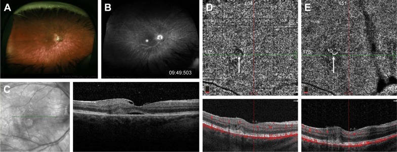
Fundus photograph of the right eye with BSCR showing multiple ovoid hypopigmented lesions in the mid-periphery (A). Late-phase fluorescein angiogram demonstrating an area of more intense hyperfluorescence at the fovea (B). Structural OCT on initial presentation showing subretinal fluid and adjacent subretinal hyper-reflective material (C). After multiple anti-VEGF injections, the subretinal fluid is resolved and, on OCT-A of the choriocapillaris, there is only a very faint area of flow signal surrounded by a halo of decreased signal (D). Twelve months later without anti-VEGF injections, OCT-A shows a clearer branching CNV network but still without any subretinal fluid (E).
Abbreviations: Anti-VEGF, anti-vascular endothelial growth factor; BSCR, birdshot chorioretinopathy; CNV, choroidal neovascularization; OCT-A, optical coherence tomography angiography.

