Figure 3.
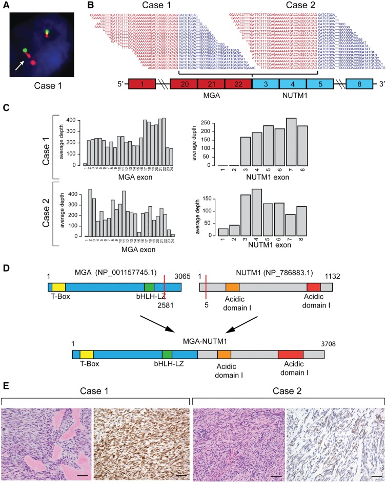
(A) Case 1: Fluorescence in situ hybridization assay showing evidence of complex NUTM1 rearrangements. The arrow indicates examples of abnormal doublet signals of the 3′ NUTM1 locus (orange). (B) RNA-sequencing reads spanning the junction between MGA exon 22 and NUTM1 exon 3. (C) RNA-seq coverage of MGA and NUTM1 exons. (D) Schematic representation of MGA and NUTM1 protein domains and resulting MGA–NUTM1 fusion protein structure. (E) H&E staining of both cases showed a monomorphic spindle cell morphology arranged in fascicles with an associated collagenous stroma. Case 1 showed dense hyalinized material, the so-called “amianthoid fibers,” resembling osteoid matrix deposition. Immunohistochemically Case 1 showed strong, diffuse nuclear reactivity for NUTM1, whereas Case 2 showed a multifocal weak staining pattern. Scale bars, 500 µM.

