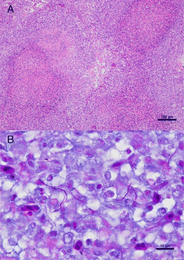
Fig. 2.

Histological sections of the lung. a Multiple granulomas with central necrosis. The granulomatous inflammation is characterized by macrophages, lymphocytes, fewer heterophils and occasional multinucleated giant cells. Hematoxylin and eosin stain (Bar = 100 μm). b Fine, beaded filamentous acid-fast organisms are visible in parts of the lesions. Fite-Faraco stain (Bar = 10 μm)
