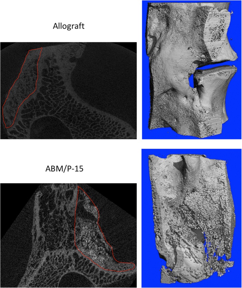
Fig. 1.

Micro-CT images showed different bone formation patterns: allograft had nice bone formation with a combination of woven and lamellar bones. ABM/P-15 also displayed nice bone formation with clearly visible unresolved residue of hydroxyapatite. Upper left: 2D section of allograft (circle), and upper right: 3D reconstruction of allograft fusion mass. Lower left: 2D section of ABM/P-15 (circle). Upper right: 3D reconstruction of ABM/P-15 fusion mass
