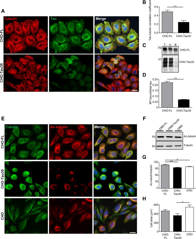
Fig. 2.
Reduced microtubule organization in CHO-Tau35 cells. a Immunofluorescence of methanol-fixed CHO-FL and CHO-Tau35 cells, labeled with antibodies to α-tubulin (red), tau (green) and Hoescht 33,342 (blue, nuclei). Scale bar = 20 μm. b Graph showing the correlation (Pearson’s) of tau colocalization with microtubules in CHO-FL and CHO-Tau35 cells. Values represent mean ± S.E.M, n = 80 cells from 3 independent experiments, Student’s t-test, **P < 0.01. c Western blots of total cell lysate (T), unbound (U), and microtubule-bound (B) fractions of CHO-FL and CHO-Tau35 cells probed with antibody to total tau. Molecular weight markers (kDa) are shown on the left. d Graph showing the ratio of tau in the microtubule (MT)-bound fraction relative to total tau in CHO-FL and CHO-Tau35 cell lysates. Values represent mean ± S.E.M., n = 3, Student’s t-test, **P < 0.01. e Immunofluorescence of paraformaldehyde-fixed CHO-FL, CHO-Tau35, and untransfected CHO cells labelled with antibodies to α-tubulin (green), acetylated α-tubulin (red) and Hoechst 33342 (blue). Scale bar = 20 μm. f Western blots of CHO-FL, CHO-Tau35 and CHO cell lysates probed with antibodies recognizing acetylated and total α-tubulin. Molecular weight markers (kDa) are shown on the left. g Graph showing the ratio of acetylated/total α-tubulin in CHO-FL and CHO-Tau35 cells, relative to CHO cells (100%). Values represent mean ± S.E.M., n = 4, one-way ANOVA, *P < 0.05, **P < 0.01. h Graph showing the areas of CHO-FL, CHO-Tau35 and CHO cells, mean ± S.E.M. A minimum of 50 cells were measured for each cell line. One-way ANOVA, *P < 0.05

