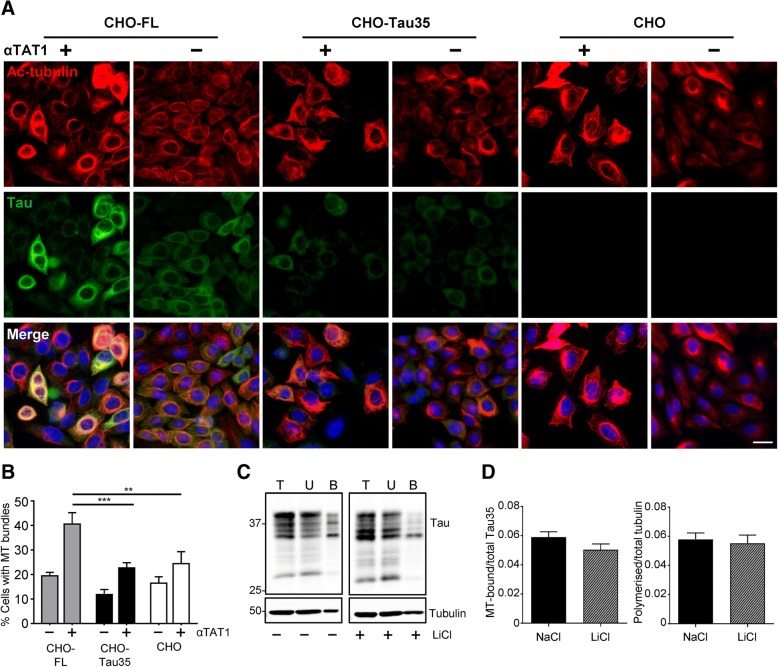
Fig. 3.
Microtubule organization in CHO-Tau35 cells is not restored by increasing tubulin acetylation or decreasing tau phosphorylation. a CHO-FL, CHO-Tau35 and CHO cells were transiently transfected with a plasmid expressing α-tubulin N-acetyltransferase 1 (αTAT1, +) or without plasmid (−). Panels show immunofluorescence of methanol-fixed cells labelled with antibodies to acetylated α-tubulin (red), tau (green), and Hoechst 33342 (blue, nuclei). Scale bar = 20 μm. b Graph showing the percentage of cells exhibiting microtubule (MT) bundles in CHO-FL, CHO-Tau35 and CHO cells in the presence (+) or absence (−) of αTAT1. Values represent mean ± S.E.M. A minimum of 150 cells were counted for each transfection condition; two-way ANOVA, **P < 0.01; ***P < 0.001. c Western blots of tau and α-tubulin in total cell lysate (T), unbound (U) and MT-bound (B) fractions of CHO-Tau35 cells treated with 5 mM LiCl (+) or 5 mM NaCl (−) for 24 h and probed with antibodies to tau or α-tubulin. d Graphs show the amount of tau present in the MT-bound fractions, relative to total Tau35 (left), and the amount of polymerized α-tubulin relative to total α-tubulin (right) in CHO-Tau35 cells, following exposure to NaCl (control) or LiCl. Values represent mean ± S.E.M., n = 3. Student’s t-test, *P < 0.05, **P < 0.01

