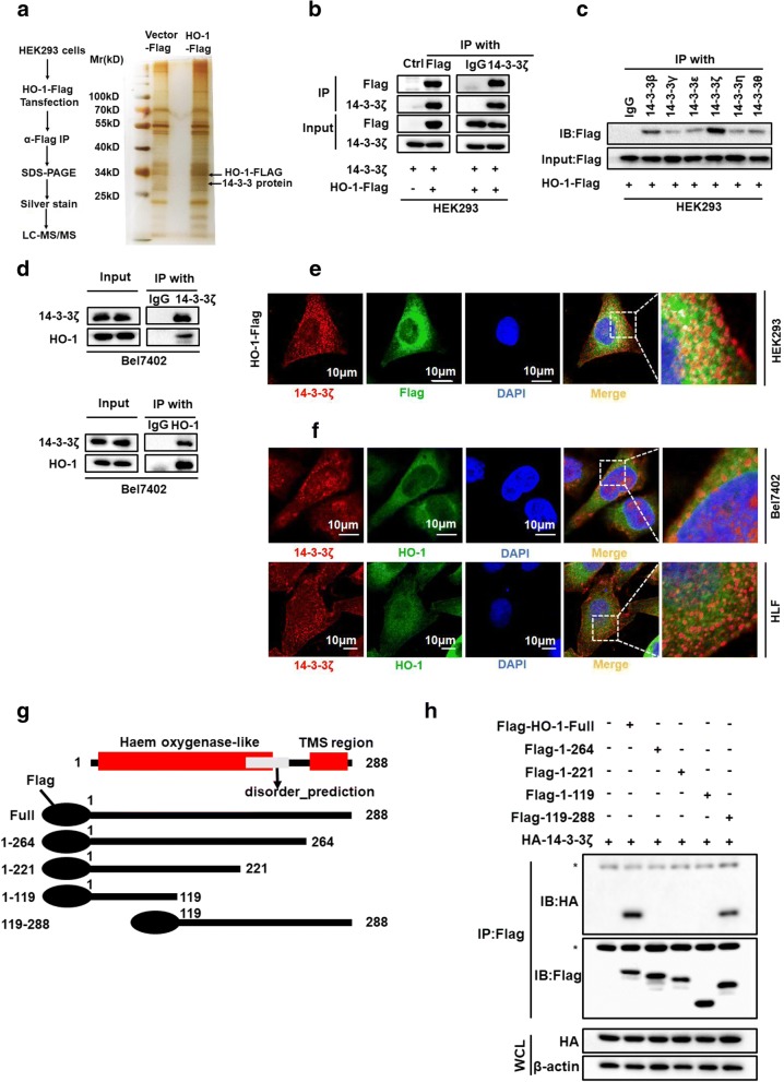
Fig. 1.
Identification of 14–3-3ζ as a binding partner for HO-1 (a) MS analysis of HO-1-associated proteins. b Whole cell lysates from HEK293 cell transfected with Flag-tagged HO-1 were subjected to co-IP with anti-FLAG or anti-14-3-3ζ antibodies and followed by immunoblotting with antibody against FLAG, 14–3-3ζ. c HEK293 cells were transfected with a plasmid encoding Flag-tagged HO-1. The lysates were immunoprecipitated with anti-14-3-3 antibodies against the indicated 14–3-3 isoforms, and were then analyzed by western blotting. d Bel7402 cell lysate was subjected to co-IP with anti-14-3-3ζ or anti-HO-1, followed by immunoblotting with antibody against 14–3-3ζ, HO-1. e HEK293 cells were transfected with Flag-tagged HO-1. After 24 h of transfection, immunofluorescence staining was performed to observe the co-localization of exogenously expressed HO-1 and 14–3-3ζ. Scale bar denotes 50 μm. f Confocal experiments were performed to determine the co-localization between endogenous HO-1 and 14–3-3ζ. Scale bar denotes 10 μm. g Schematic diagram of FLAG-tagged full-length or deletion constructs of HO-1 used in this study. h Whole cell lysate from HEK293 cell transfected with the indicated plasmids were subjected to co-IP with anti-FLAG and followed by immunoblotting with indicated antibodies. The asterisk denotes a nonspecific band. WCL denotes whole-cell lysate

