. 2018 Dec 10;114:e180438. doi: 10.1590/0074-02760180438

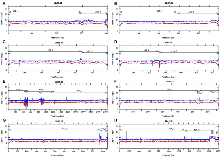
Fig. 2: read-depth analysis throughout the chromosomes formed by the union of two or more PacBio-assembled contigs. Coverage (log2 value) was appointed by sliding window analysis (bin 200 bp) with either Illumina (in blue) and PacBio (in red) reads, along chromosomes 2, 8, 9, 10, 20, 22, 27 and 34. Contigs lengths are shown by arrowheads lines. Chromosomes 2, 20, and 22 (panels A, E and F) were joined using the minimus 2 assembler. Chromosomes 8 (panel B) and 9 (panel C) were joined by the SSPACE-standard tool. Moreover, chromosome 27 (panel G) was joined by the SSPACE-LongRead tool and chromosome 10 (panel D) was joined with the pyScaf scaffolder tool. Finally, pyScaf and minimus2 were necessary for joining chromosome 34 (panel H).

Fig. 2: