Figure 7.

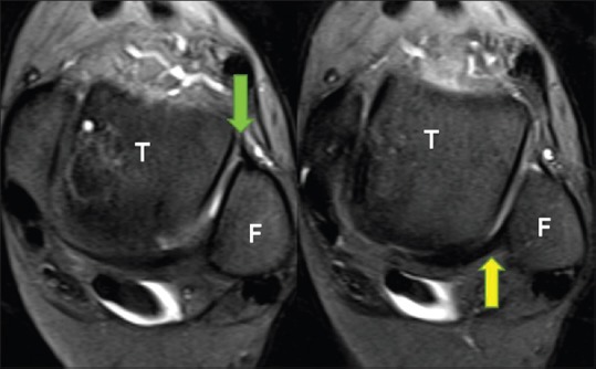
Normal anterior (green arrow) and posterior (yellow arrow) inferior tibiofibular or syndesmotic ligaments are best identified on axial images, where the talar dome (T) is square shaped, and Fibula is round

Normal anterior (green arrow) and posterior (yellow arrow) inferior tibiofibular or syndesmotic ligaments are best identified on axial images, where the talar dome (T) is square shaped, and Fibula is round