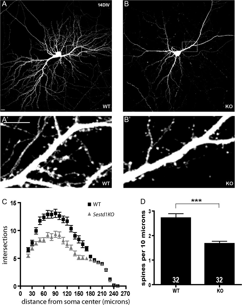
Figure 1.
Sestd1 mutant neurons have reduced dendrite arbors and spines. (A,B) Cultured hippocampal neurons (HCNs) transfected with GFP and imaged at 14 DIV. (A) WT (B) Sestd1KO. (A′,B′) Corresponding boxed regions from A to B at higher magnification. (C) Sholl analysis of WT (black squares) and Sestd1KO (gray triangles) HCNs. (D) Spine density is reduced in Sestd1KO HCNs. Scale bars: 10 μm. The number within each bar = n, 32 neurons/condition. ***P ≤ 0.001.

