Figure 5.
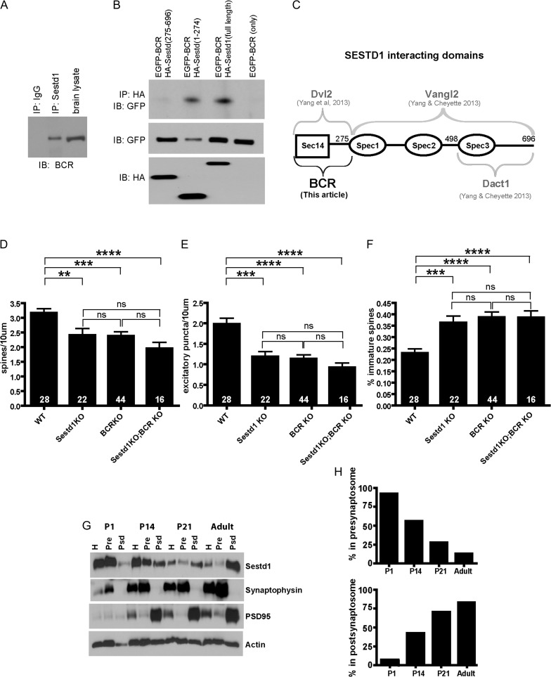
Sestd1 forms physiological complexes with BCR and has a developmentally dynamic presynaptic versus postsynaptic distribution. (A) Adult hippocampal lysate (wild type, C57Bl/6 J) immunoprecipitated with anti-Sestd1 antibody; co-precipitated BCR detected with anti-BCR antibody. (B) HA-tagged full-length Sestd1 and deletion mutants tested for ability to pull down ECFP-tagged BCR when the proteins are recombinantly expressed in HEK293T cells. (C) Schematic representation of Sestd1 domains contributing to interaction with BCR (this study), compared with similar determinations for domains contributing to interactions with Dvl2, Vangl2 and Dact1 (prior studies, Cheyette lab). (D–F) Deletion of either Sestd1 or BCR alone produce similar phenotypic effects with regard to dendritic spine density (D), excitatory synapse density (E), and spine maturity (F), in cultured forebrain neurons. Phenotypic effects of simultaneous Sestd1 and BCR loss are not significantly different from effects resulting from the single mutants. The number within each bar = n, animals/genotype. ns, P > 0.05; **P ≤ 0.01; ***P ≤ 0.001, ****P ≤ 0.0001. (G) Sestd1 synapse localization shifts during postnatal development. Presynaptic and postsynaptic compartment proteins were separated in a brain lysate from a WT (CD-1) mouse using biophysical techniques and the distribution of Sestd1 determined by immunoblotting. Synaptophysin and PSD95 were used as presynaptic and postsynaptic markers respectively for the quality of biophysical separation of the desired synaptosome fractions. (H) Quantification of distribution of Sestd1 into presynaptic and postsynaptic biophysical fractions at different time points.

