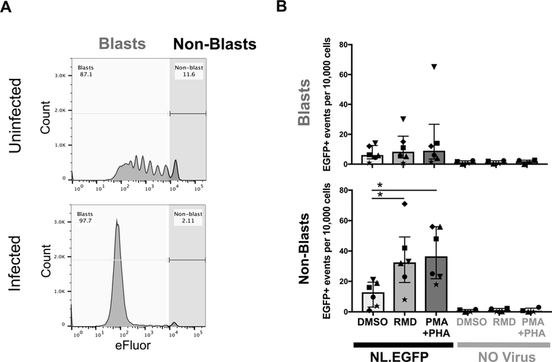
Fig. 4. Post-activation latency following infection with NL4.3EGFP.

A. Activated CD4+ T cells were infected with NL4.3-EGFP virus and cultured for 12 days to allow cells to return to rest. Cells were sorted at day twelve post-infection for EGFP-negative CD69−CD25−HLA-DR− transitional blasts (eFluorlow) and non-blasts (eFluorhigh) as shown. B. Sorted blasts and non-blasts were reactivated with the HDACi romidepsin (RMD), PMA/PHA or with control DMSO. The number of EGFP+ cells per 10,000 events was quantified by flow cytometry. In each experiment between 10,000 and 100,000 events were collected. Data represent mean ± SEM of six individual donors. Each symbol represents a different donor. * p<0.05
