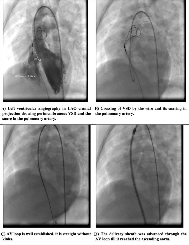
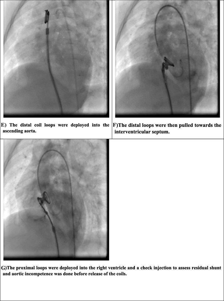
Figure 1.
The procedure of VSD closure. (A) Left ventricular angiography in LAO cranial projection showing perimembranous VSD and the snare in the pulmonary artery. (B) Crossing of VSD by the wire and its snaring in the pulmonary artery. (C) AV loop is well established; it is straight without kinks. (D) The delivery sheath was advanced through the AV loop until it reached the ascending aorta. (E) The distal coil loops were deployed into the ascending aorta. (F) The distal loops were then pulled toward the interventricular septum. (G) The proximal loops were deployed into the right ventricle and a check injection to assess residual shunt and aortic incompetence was done prior to release of the coils. AV = arteriovenous; LAO = left anterior oblique; VSD = ventricular septal defect.


