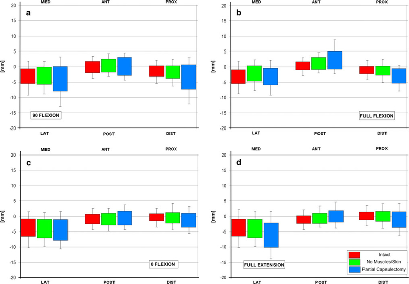
Fig. 2.
Displacements of the femoral head relative to the acetabulum in medial/lateral (MED/LAT), anterior/posterior (ANT/POST) and proximal/distal (PROX/DIST) during each test and hip position with each histogram representing the testing in different hip flexion– extension positions, and each different colour bar representing the different soft tissue states (I intact, II after removal of skin and muscles, while leaving the capsuloligamentous structures intact, and III after partial circumferential capsulectomy). a Femoral head motion in the 3 planes while the hip was in 90 degrees of flexion and the hip rotated and abducted–adducted. b is for the same measurements while the hip was in full flexion, c in neutral flexion– extension and d with the hip in hyperextension

