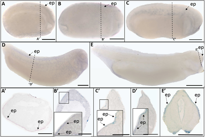
Fig. 5.

Whole mount (A-E) and histological (A’-E’) expression of trpv6 transcripts in X. laevis embryos. Lateral view for all whole mount embryos, anterior to the right; dorsal to the top for all histology images. (A, A’) stage 15 (mid-neurula stage); (B, B’) stage 20 (late neurula stage); (C, C’) stage 25 (early tailbud stage); (D, D’) stage 30 (late tailbud stage); (E, E’) stage 35 (swimming tadpole stage). Arrows indicate regions of gene expression (ep, epidermis). Dashed lines represent positions of corresponding sections. Scale bars = 250µm.
