Fig. 1.
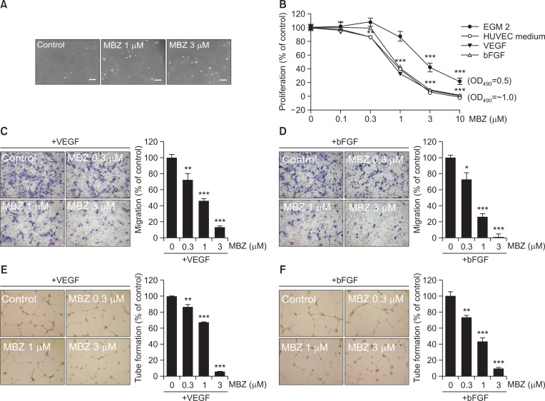
Mebendazole inhibits proliferation, migration and tube formation of ECs. (A) HUVECs were treated with MBZ (1, 3 μM) for 24 h, and observed by light microscopy. Scale bars: 100 μm. (B) HUVECs were treated with the indicated concentrations of MBZ in several medium conditions including EGM2, HUVEC medium, and M199 containing VEGF (10 ng/ml) or bFGF (10 ng/ml). After 48 h, MTS assay was performed. The percentage of proliferation was calculated based on cell proliferation of respective indicated culture medium control group. (C, D) HUVECs pre-treated with MBZ for 30 min were allowed to migrate into bottom chamber with VEGF (5 ng/ml) or bFGF (5 ng/ml) for 5 h. Then, the migrated cells were counted per view field. (E, F) Tube formation of HUVECs pre-treated with MBZ for 30 min on Matrigel was performed in the presence of VEGF (10 ng/ml) or bFGF (10 ng/ml) for 12 h. The mean of total capillary tube length per view field was measured. *p<0.05, **p<0.01, and ***p<0.001 vs. control (MBZ 0 μM) group.

