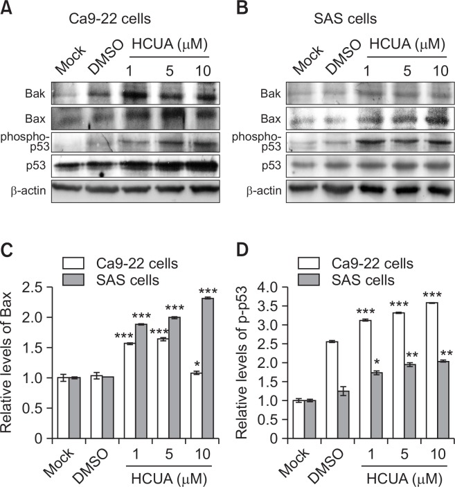
Fig. 4.
Relative protein of pro-apoptotic genes in cis-3-O-p-hydroxy-cinnamoyl ursolic acid (HCUA)-treated cells. The protein levels of Bak, Bax, phospho-p53 (Ser15), and p53 in Ca9-22 (A) and SAS (B) cells treated with HCUA were characterized 48 h post-treatment by western blot analysis. The intensity of each immuno-reactive band for Bax or phospho-p53 (Ser15) was quantified using Image J; their relative intensities were normalized by β-actin (C, D), respectively. *p<0.05, **p<0.01, ***p<0.001 compared with mock cells.

