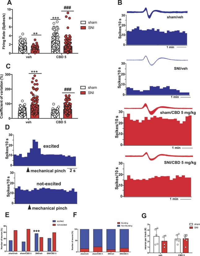
Figure 5.
Repeated CBD treatment prevents SNI-induced alterations in DRN 5-HT neuronal activity. (A) Mean DRN 5-HT firing activity (spikes/s). (B) Representative firing rate histograms of the mean DRN 5-HT firing rate activity recorded in sham and SNI rats treated with veh or CBD (5.0 mg/kg/day, for 7 days, subcutaneously [s.c.]). (C) Mean DRN 5-HT COV (%). Each bar represents mean ± SEM. Two-way ANOVA followed by Bonferroni post hoc comparisons. (D) Representative rate histograms recorded from a DRN neuron excited (top) or not (bottom) by mechanical pinch to the operated hind paw. (E) Contingency interleaved bars showing percentage (%) of DRN 5-HT neurons excited or not by mechanical paw pinch stimulation. (F) Contingency stacked bars showing percentage (%) of bursting and nonbursting DRN 5-HT neurons (the χ2 test). (G) Mean number of DRN 5-HT neuron recorded per electrode descent. Sham rats treated with veh (n = 5) or CBD (5 mg/kg/day, s.c., for 7 days) (n = 5) and SNI rats treated with veh (n = 4) and CBD (5 mg/kg/day, s.c., for 7 days) (n = 7) were tested. Two-way ANOVA followed by Bonferroni post hoc comparisons. In A and C, each point represents a single neuron recorded in each group. In F, each point represents data from an individual rat. **P < 0.01, ***P < 0.001 vs sham/veh., #P < 0.05, and ###P < 0.001 vs SNI/veh. ANOVA, analysis of variance; CBD, cannabidiol; COV, coefficient of variation; DRN, dorsal raphe nucleus; SNI, spared nerve injury.

