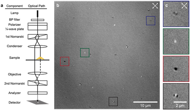
Figure 1. Wide-field DIC microscopy of AuNS.
(a) Scheme of DIC optical components and light path. (b) Wide-field DIC image of AuNS dispersed on the coverslip. (c) Zoomed-in DIC images of particles in colored squares highlight the distinct patterns produced by different AuNS. White arrows indicate the polarizations of the ordinary and extraordinary beams.

