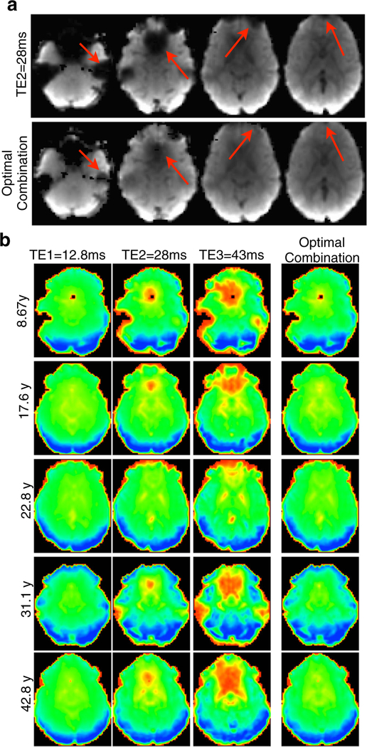
Fig. 3.
Signal dropout recovery after optimal combination of multi-echo time series datasets across the age range. a shows the benefit of using an optimally combined slice stack over a typical slice stack of an EPI volume collected at an echo time of 28ms, with arrows pointing to significant areas of signal improvement. b further displays the improvement in EPI quality by showing slices in ventral regions at three echo times and the action of the weighting filter in combining the echoes at those slices, compensating for signal dropout in these regions for datasets of subjects across the age range

