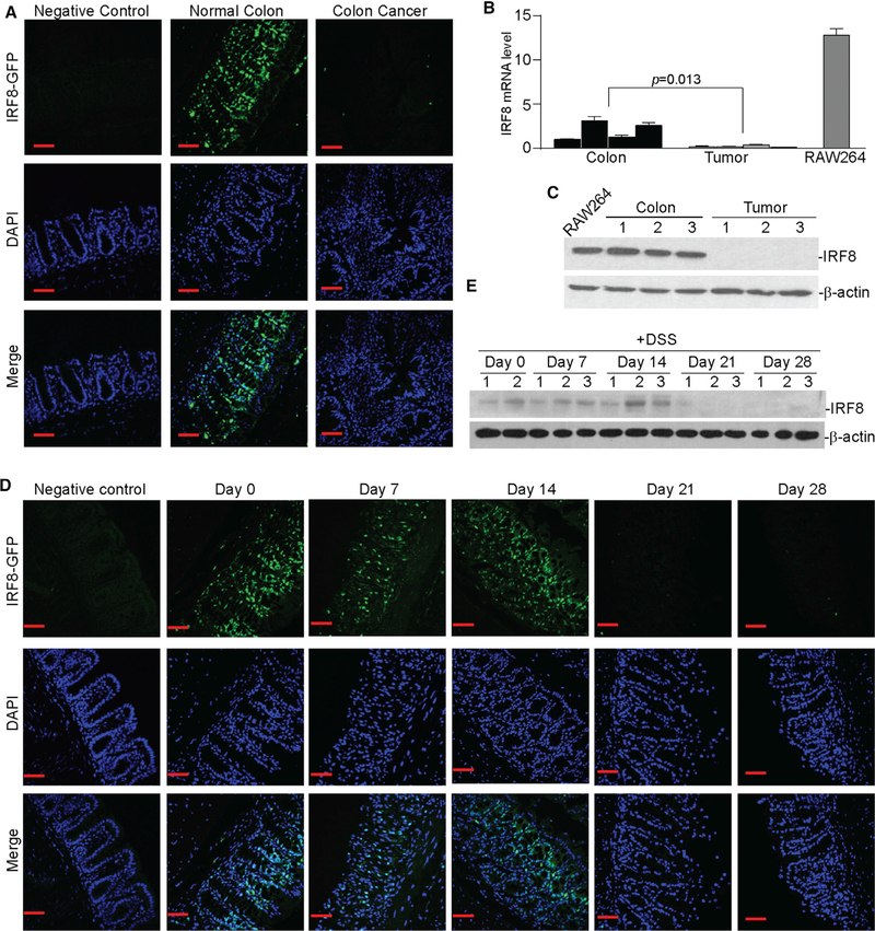
Figure 2. Chronic Inflammation Silences IRF8 Expression in Colonic Epithelial Cells.
(A) IRF8-GFP reporter mice were treated with the AOM-DSS cycle to induce colitis-associated colon cancer. Colon tissues from normal (negative control, n = 3), untreated IRF8-GFP reporter (normal colon, n = 3), and AOM-DSS-treated GFP-reporter (colon cancer, n = 3) mice were fixed and frozen. The frozen sections were stained with DAPI and analyzed for GFP fluorescence intensity using a confocal microscope. Shown are representative images. Scale bar, 50 μm.
(B) RNA was extracted from the colon tissues of tumor-free (colon, n = 4) and tumor-bearing (tumor, n = 4) mice and cultured RAW264 cells and analyzed for IRF8 mRNA expression levels by qPCR using IRF8-specific primers. β-Actin was used as an internal control. Each column represents the mean of triplicates from one mouse. Four mice were included in each group. Bar, SD. RAW264 was used as a positive control for IRF8 expression.
(C) Colon tissues of tumor-free (colon, n = 3) and tumor-bearing (tumor, n = 3) mice and cultured RAW264 cells were lysed and analyzed by western blotting analysis using IRF8-specific antibody. β-Actin was used as a normalization control.
(D) IRF8-GFP reporter mice were treated with DSS in drinking water for 7 days, followed by drinking water for 14 days, then DSS again for 7 days. Colon tissues were collected at days 0, 7, 14, 21, and 28 and analyzed for GFP fluorescence intensity. Normal mice were used as negative controls. Scale bar, 50 μm.
(E) Total protein lysate was prepared from the colon tissue of mice as in (C) at days 0, 7, 14, 21, and 28 and analyzed for IRF8 protein level by western blotting. β-Actin was used as a normalization control.

