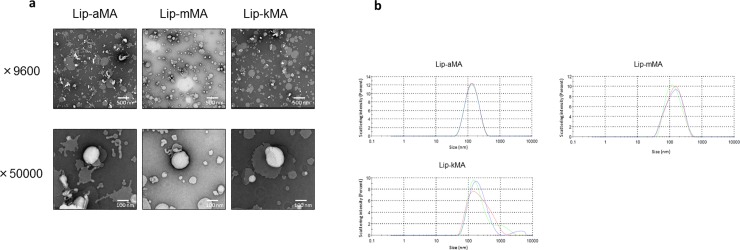
Fig 2.
a, b: Electron micrographs of liposomal MA suspension and the particle distribution. (a) Lip-aMA was heteromorphic and seemed to be partially disrupted. Lip-mMA and Lip-kMA showed generally round and flat edges. (b) The particle distribution of Lip-aMA, Lip-mMA and Lip-kMA demonstrated by laser-Doppler velocimetry with a ZEN3600.

