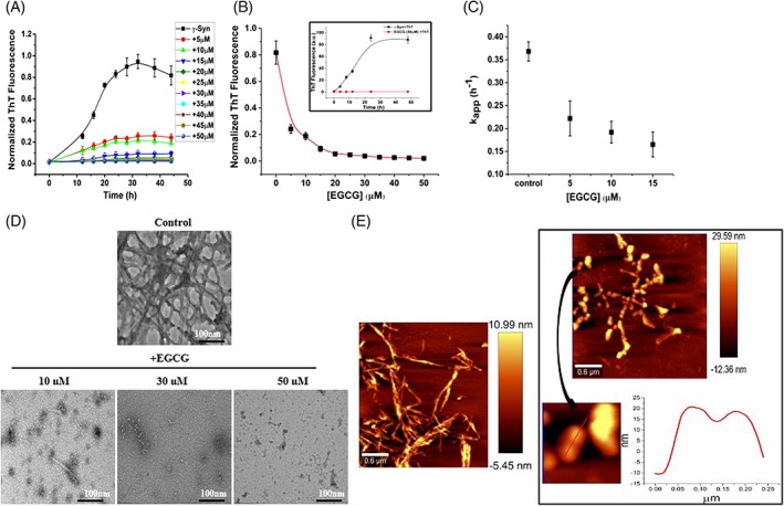
Figure 1.

EGCG suppresses γ‐Syn fibrillation in a concentration‐dependent manner. Fibrillation kinetics of γ‐Syn (75 μM) studied using thioflavin T (ThT) assay in the presence of increasing concentration of EGCG (5–50 μM). (A) Decrease in ThT fluorescence with respect to time of fibrillation and (B) with respect to EGCG concentration, (inset) control of ThT and EGCG showing absence of interference between the two. The error bars represent the standard deviation from three independent aggregation reactions, ±SD (n = 3). (C) The apparent rate of fibrillation (kapp) is shown to decrease with an increasing concentration of EGCG. D) Transmission electron microscopy images of γ‐Syn fibrils formed at the end of fibrillation in the presence of increasing concentration of EGCG (Scale bars, 100 nm). E) Atomic force microscopy images showing significant disappearance of mature γ‐Syn fibrils in the presence of EGCG (50 μM) with an appearance of self‐associated large spherical oligomers clearly depicted in the cross section of the oligomers (right panel; below).
