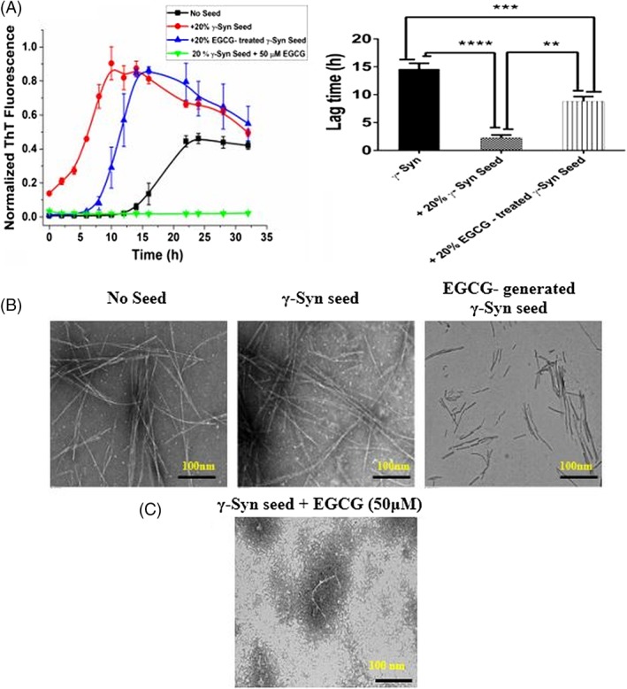
Figure 4.

EGCG inhibits γ‐Syn elongation and forms kinetically retarded on‐pathway oligomers: (A) (Left panel) Fibrillation kinetics of γ‐Syn in the presence of EGCG (50 μM) treated and untreated seeds (20% v/v) shows an increased lag time of fibrillation in the presence of EGCG generated seeds as compared to the untreated γ‐Syn seeds. Negligible ThT fluorescence in the presence of EGCG (50 μM) added to the seeded γ‐Syn fibrillation (green line) shows elongation inhibition. The data have been normalized with respect to the maximum fluorescence intensity recorded in the seeded γ‐Syn and the error bars represent ±SD (n = 3). (Right panel) Bar graph showing the significant difference in the lag time in the EGCG‐generated seeds as compared to the seeds formed without EGCG (**P < 0.001) and between non‐seeded γ‐Syn and EGCG‐generated γ‐Syn seeds (***P < 0.0005). The lag time of fibrillation in the presence of untreated γ‐Syn seeds was significantly shorter than the non‐seeded kinetics (****P < 0.0001). (B) The TEM images showing the morphology of the fibrils formed (left to right: no seed, γ‐Syn seed, EGCG‐treated γ‐Syn seeds) (Scale bars, 100 nm). (C) TEM image showing formation of short, broken fragments upon addition of EGCG (50 μM) to seeded γ‐Syn pathway indicating inhibition of fibril elongation by EGCG.
