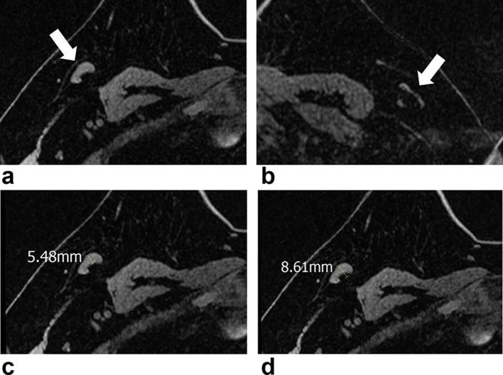
Figure 3.
MR images of a 47-year-old female with breast cancer in her right breast. Axial non-enhanced, fat-suppressed T1 weighted images (a–d) show ipsilateral (a, arrow) and contralateral (b, arrow) ALNs. Ipsilateral ALN had a cortical thickness of 5.5 mm (c) and its short diameter was 8.6 mm (d). The contralateral ALN (d) had a cortical thickness of 1.6 mm and its short diameter was 3.4 mm. Ultrasound-guided aspiration of the ipsilateral ALN with subsequent surgical resection revealed nodal metastases. ALN, axillary lymph node.

